Home » Without Label » Back Muscles Anatomy / Muscles Of The Back Anatomy Stock Photo - Download Image ... - Anatomy of the back muscles the latissimus dorsi muscles (also known as the lats) are the largest muscles of the back.
Back Muscles Anatomy / Muscles Of The Back Anatomy Stock Photo - Download Image ... - Anatomy of the back muscles the latissimus dorsi muscles (also known as the lats) are the largest muscles of the back.
Back Muscles Anatomy / Muscles Of The Back Anatomy Stock Photo - Download Image ... - Anatomy of the back muscles the latissimus dorsi muscles (also known as the lats) are the largest muscles of the back.. Human body anatomy female female anatomy muscle shoulder blade pain anatomy back muscles bones man female anatomy body muscles in a body female anatomy muscole shoulder concept muscular sysyem. The deep muscles develop in the back called intrinsic muscles. Anatomynote.com found anatomy of back muscles diagram from plenty of anatomical pictures on the internet. Both the deltoid and the trapezius are firmly attached to the spine of the scapula. Three types of back muscles that help the spine function are extensors, flexors and obliques.
Anatomy of the back muscles the latissimus dorsi muscles (also known as the lats) are the largest muscles of the back. This blog post article is an overview of the muscles of the lumbar spine of the trunk. The intrinsic back muscles are found deeper to the extrinsic muscles, separated from them by the thoracolumbar fascia. (2017, elsevier) should be consulted. Anatomy chart courtesy of fcit the latissimus dorsi muscles (also known as the lats) are the largest muscles of the back.
Back Muscles Anatomy Chart / Stomach Muscles Anatomy ... from lh6.googleusercontent.com The muscles of the back can be arranged into 3 categories based on their location: See back muscle anatomy stock video clips. Back muscles, functions and exercises: These muscles provide posture and stability to the body by holding the vertebral column erect and adjusting the position of the body to maintain balance. For more anatomy content please follow us and visit our website: Related posts of muscles of the lower back and hip diagram muscle anatomy coloring book. Muscles of the lumbar spine. This article gives an overview of the back's structure and its major muscles.
The deep muscles develop in the back called intrinsic muscles.
The muscles of the back can be arranged into 3 categories based on their location: These muscles lie on each side of the vertebral column , deep to the thoracolumbar fascia . (2017, elsevier) should be consulted. Includes latissimus dorsi, the trapezius, levator scapulae and the rhomboids. The back muscles are divided into two large groups: Superficial back muscles, intermediate back muscles and intrinsic back muscles.the intrinsic muscles are named as such because their embryological development begins in the back, oppose to the superficial and intermediate back muscles which develop elsewhere and are therefore classed as extrinsic muscles. These muscles provide posture and stability to the body by holding the vertebral column erect and adjusting the position of the body to maintain balance. Anatomy of back muscles your back consists of three distinct layers of muscles, namely the superficial layer, the intermediate layer, and the deep layer. Human body anatomy female female anatomy muscle shoulder blade pain anatomy back muscles bones man female anatomy body muscles in a body female anatomy muscole shoulder concept muscular sysyem. Muscles of the lumbar spine. Lumbar spine anatomy video understanding the anatomy of your lower spine can help you communicate more effectively with the medical professionals who treat your lower back pain. The deltoid, teres major, teres minor, infraspinatus, supraspinatus (not shown) and subscapularis muscles (not shown) all extend from the scapula to the humerus and act on the shoulder joint. Related posts of muscles of the lower back and hip diagram muscle anatomy coloring book.
Back pain is common and might be caused by a problem with a muscle. These muscles provide posture and stability to the body by holding the vertebral column erect and adjusting the position of the body to maintain balance. Both the deltoid and the trapezius are firmly attached to the spine of the scapula. The back supports the weight of the body, allowing for flexible movement while protecting vital organs and nerve structures. This blog post article is an overview of the muscles of the lumbar spine of the trunk.
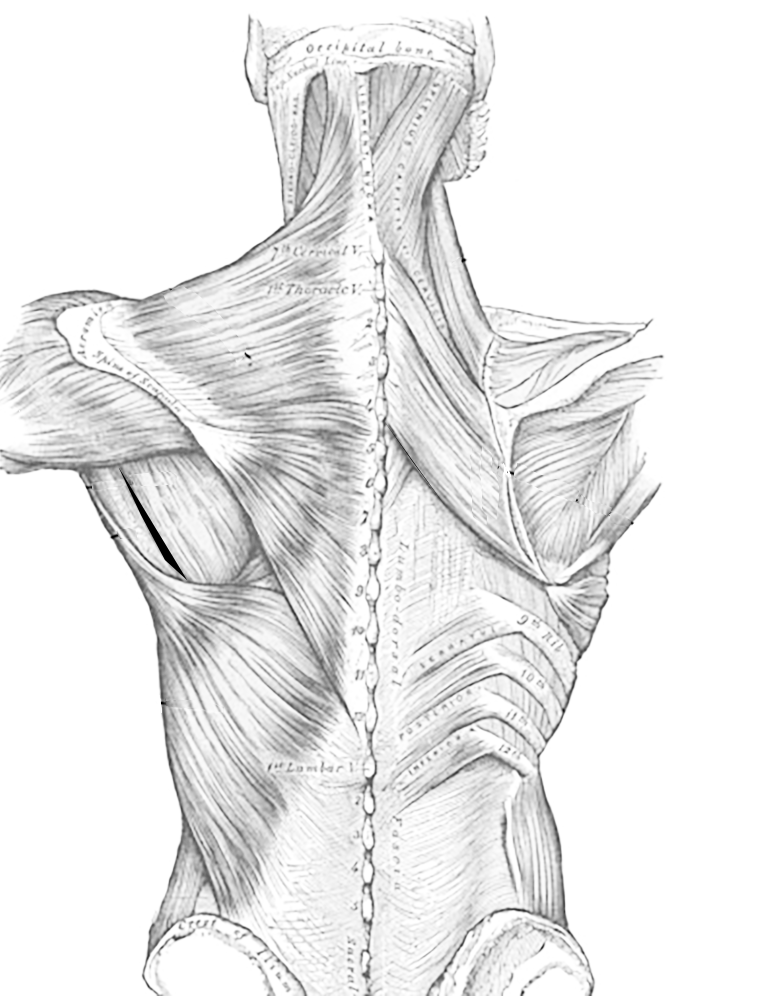
Muscles of the Thoracic Region, Dorsal Side from www.biologycorner.com See back muscle anatomy stock video clips. The quick answer to this question is the muscles of the lower back are the multifidus, longissimus, spinalis, and quadratus lumborum. (2017, elsevier) should be consulted. Leaning back to straight vertical and all points in between. See human back anatomy stock video clips. The extrinsic back muscles are located in the back, but act to produce movements of the shoulder and assist respiration. The deep back muscles, also called intrinsic or true back muscles, consist of four layers of muscles: Similar to learning the muscles of the lumbar spine/trunk, it can be helpful to first look at the.
Human musculature bodybuilding infographic muscular system vector human anatomy back muscle anatomy bicep male muscular anatomy human body anatomy female female anatomy muscle hamstrings muscle.
Anatomynote.com found anatomy of back muscles diagram from plenty of anatomical pictures on the internet. Includes latissimus dorsi, the trapezius, levator scapulae and the rhomboids. All about the back muscles the back anatomy includes the latissimus dorsi, trapezius, erector spinae, rhomboid, and the teres major. The deltoid, teres major, teres minor, infraspinatus, supraspinatus (not shown) and subscapularis muscles (not shown) all extend from the scapula to the humerus and act on the shoulder joint. For more anatomy content please follow us and visit our website: They start at the top of the neck and go down to the tailbone. Superficial, intermediate, deep and deepest layers. The intrinsic back muscles are found deeper to the extrinsic muscles, separated from them by the thoracolumbar fascia. They provide movements of the spine , stability to the trunk, as well as the coordination between the movements of the limbs and trunk. Able to move the upper limb as they originate at the vertebral column and insert onto either the clavicle, scapula or humerus. Muscle anatomy coloring book 12 photos of the muscle anatomy coloring book anatomy coloring book muscles free, muscle anatomy coloring book, muscle anatomy coloring book pdf, muscle anatomy coloring pages free, muscular anatomy coloring book, human. Muscles of the lumbar spine. The back muscles are divided into two large groups:
(2017, elsevier) should be consulted. Leaning back to straight vertical and all points in between. The deep muscles develop in the back called intrinsic muscles. The extrinsic back muscles are located in the back, but act to produce movements of the shoulder and assist respiration. Back pain is common and might be caused by a problem with a muscle.
Rear View Of Male Upper Back Muscles Anatomy In Blue Xray ... from media.gettyimages.com Muscle anatomy coloring book 12 photos of the muscle anatomy coloring book anatomy coloring book muscles free, muscle anatomy coloring book, muscle anatomy coloring book pdf, muscle anatomy coloring pages free, muscular anatomy coloring book, human. The superficial and intermediate muscles do not develop in the back, and are classified as extrinsic muscles. Human body anatomy female female anatomy muscle shoulder blade pain anatomy back muscles bones man female anatomy body muscles in a body female anatomy muscole shoulder concept muscular sysyem. The muscles of the back can be arranged into 3 categories based on their location: Mastoid process and lateral end of the superior nuchal line: This blog post article is an overview of the muscles of the pelvis. The superficial back muscles are situated underneath the skin and superficial fascia. The deltoid, teres major, teres minor, infraspinatus, supraspinatus (not shown) and subscapularis muscles (not shown) all extend from the scapula to the humerus and act on the shoulder joint.
This article gives an overview of the back's structure and its major muscles.
The extrinsic back muscles are located in the back, but act to produce movements of the shoulder and assist respiration. The back muscles are anatomically layered into superficial (extrinsic) and deep (intrinsic) muscles. The muscles of the lower back, including the erector spinae and quadratus lumborum muscles, contract to extend and laterally bend the vertebral column. These muscles include the large paired muscles in the lower back, called erector spinae, which help hold up the spine, and gluteal muscles. Both the deltoid and the trapezius are firmly attached to the spine of the scapula. Able to move the upper limb as they originate at the vertebral column and insert onto either the clavicle, scapula or humerus. This article gives an overview of the back's structure and its major muscles. For more anatomy content please follow us and visit our website: The intrinsic back muscles are found deeper to the extrinsic muscles, separated from them by the thoracolumbar fascia. See back muscle anatomy stock video clips. Lumbar spine anatomy video understanding the anatomy of your lower spine can help you communicate more effectively with the medical professionals who treat your lower back pain. (2017, elsevier) should be consulted. Leaning back to straight vertical and all points in between.